Friday, April 27, 2012

Triceps tear

1-Less common than biceps tear.

2-Anatomy


1, Ulna. 2, Radial head. 3, Lateral epicondyle. 4, Extensor carpi radialis longus muscle. 5, Olecranon fossa. 6, Medial epicondyle. 6a, Common flexor tendon. 7, Triceps muscle (lateral head). 8, Humerus. 9, Triceps muscle (Medial head). 10, Flexor digitorum superficialis muscle. 11, Supinator muscle. 12, Extensor digitorum muscle.
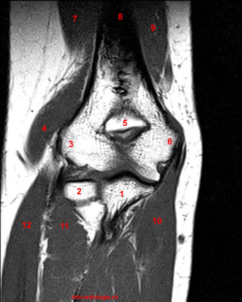
Wednesday, April 25, 2012

Biceps injury

1-It is a clinical diagnosis.

2-Tendon will be corrugated and interrupted.


young male hand pain and weakness while work out in Gymnasium. Patient heard a snapping sound from elbow and reported to nearby doctor who advised MRI after which patient was given above elbow slab. MRI was reported as distal biceps rupture with inflammation surrounding area.

3-FABS position:

-Here the patient lies prone with the affected arm raised beside his head and the elbow is flexed passing above the head.

Localizer MR image with lines shows slice positioning for flexed abducted supinated view. Notice sections, sagittal to long axis of body but coronal to anatomy at elbow. Ideal angulation is planned along distal biceps brachii tendon, but often, as here, this structure is not clearly visible on localizer images. In this case, sections nearly perpendicular to radius provide reasonable and reproducible imaging plane.


Photograph shows patient positioning for flexed abducted supinated view: patient is positioned prone on MRI table with elbow in flexed abducted supinated view position. Notice position of arm, flexed at elbow and abducted at shoulder with supinated forearm, thumb up.

 In general, it was preferable for the patient to lie prone for these views. The shoulder was abducted 180°, with the arm beside the head. The elbow was flexed to 90°, with the forearm supinated, thumb up, and a shoulder phased array coil was placed around the elbow . The position is referred to in this article as the flexed abducted supinated view, but usually in our practice it is termed the “FABS view,” meaning the flexed elbow with the shoulder abducted and the forearm in supination view.


Fast spin-echo proton-density–weighted MR image (repetition time msec/echo time msec = 3000/34) obtained with the patient in the FABS position shows a normal distal biceps tendon (curved arrow), the musculotendinous junction (straight arrow), and the radial tuberosity (arrowhead).


 Fat-suppressed fast spin-echo proton-density–weighted MR image (3000/45) demonstrates a minor partial tear of the distal biceps tendon (arrow) with a trace of peritendinous fluid (arrowhead).


Golfer's elbow or medial epicondilitis

1-Degeneration of the common flexor tendon secondary to overload of the flexor muscle group that arises from the medial epicondyle resulting in

-Muscle strain injury---Appears as intermediate to hyper intense signal within muscle tissue but less than that of the fluid.

-Tendon degeneration (tendinosis).

-Tendon disruption.

Tuesday, April 24, 2012

Tennis Elbow

1-95% of patients occurs in non tennis players.

2-It is due to lateral epicondylitis.

3-It is the most common soft tissue abnormality affecting elbow joint.

4-Lateral epicondylitis is an over use syndrome due to repeated micro trauma of the site of origin of the common extensors tendon.


5-So it is not an inflammation or due to tennis, it is due to small tear in the tendon.

6-It could be appears as sprain or tear as mentioned before.

7-Tear has a fluid signal more than sprain.

8-Sprain in T1 has intermediate signal while in T2 it has more higher signal but less than that of the fluid.

Coronal (A) and axial (B) fat-suppressed T2-weighted images demonstrate T2-hyperintensity compatible with an undersurface partial tear of the common extensor tendon origin (arrows). The radial collateral ligament (arrowhead) is mildly thickened but intact +joint effusion.

Radial collateral ligament pathology

The same as ulnar collateral ligament (sprain and tear which could be partial or complete).


Normal anatomy: T2-weighted fat-suppressed coronal (C), T1-weighted coronal (D), and T2-weighted axial (E) images demonstrate the common extensor tendon (arrows) as low signal intensity on T2 and T1-weighted images. The radial collateral ligament (arrowheads) is seen deep to the common extensor tendon.


Lateral collateral ligament injury

Ulnar collateral ligament injury

1- A common injury.

2-Types of injury:

-Sprain:

*The ligament should has continuity but with increased thickness and or hazy outline or bright signal (this definition is applied all through the body).

-Tears:

*When the ligament shows discontinuity which could be partial or complete.

-It is not essential to differentiate between sprain and partial tear as usually they have almost the same line of treatment.

-It is essential to differentiate between partial and complete tear as the latter could have interventional line of treatment.

3-Common flexor tendon is common to be injured with ulnar collateral ligament.

4-It is essential to locate the site of the tear if it is ulnar side, humeral side or mid tear (most common).


Coronal STIR image (TR/TE, 6,000/30; inversion time, 150 milliseconds) depicts complete tear at humeral insertion (arrow) of lateral ulnar collateral ligament. Capitellar marrow edema (asterisk) is also noted.

Sunday, April 22, 2012

Structures seen in each view

Coronal view:

Lateral side:

1-Radial collateral ligament proper inserted in radius and lateral ulnar collateral proper inserted in ulna.

2-Common extensors tendon.

Medial side:

1-Medial epicondyle which is more prominent than lateral epicondyle.

2-Anterior bundle of the ulnar collateral ligament(from medial epicondyle to ulna).

3-Common flexor tendon.

Sagittal view:

1-Here you can see muscles and tendons.


Axial view:

1-


Above the elbow the radial nerve (arrow) lies between the brachioradialis (Brd) and brachialis muscles (Br) and is typically outlined by a small layer of fat on axial T1-weighted images. The biceps (B) and extensor carpi radialis longus (ECRL)muscles are also indicated.


An axial T1-weighted image just above the elbow joint demonstrates that the radial nerve has bifurcated into the superificial radial nerve (arrowhead) and posterior interosseous nerve (arrow). The brachioradialis (Brd), brachialis (Br), the extensor carpi radialis longus (ECRL) muscles and the biceps tendon (B) are indicated.


An axial T1-weighted image distal to the radiocapitellar joint demonstrates the posterior interosseous nerve (arrow) between the superficial (Ss) and deep (Sd) heads of the supinator muscle. The superficial radial nerve (arrowhead) courses between the supinator and brachioradialis muscles into the distal forearm.


The posterior interosseous nerve (arrowhead) is identified between the deep head of the supinator (Sd) and the tendinous proximal edge of the superficial head of the supinator muscle (arcade of Frohse) (arrow).

Upper limb nerves


Saturday, April 21, 2012

Muscles

1-We have four groups.

2-Anterior group:

-Formed from biceps and brachialis.

Above the elbow the radial nerve (arrow) lies between the brachioradialis (Brd) and brachialis muscles (Br) and is typically outlined by a small layer of fat on axial T1-weighted images. The biceps (B) and extensor carpi radialis longus (ECRL)muscles are also indicated.

3-Posterior group:

-Formed from triceps and anconeus.


Normal triceps muscle and tendon. A, Sagittal T1-weighted MR image shows the triceps muscle (M) and its tendon (white arrows) inserting on the olecranon (O). Fat (F) is present in the olecranon fossa. The box delineates the field of view in the corresponding sonographic image.


4-Medial groups=flexors muscles arises from medial condyle as common tendon= pronator teres and palmaris longus and flexors.

5-Lateral groups=extensors muscles arise from lateral epicondyle
as common tendon.

Radial collateral ligament

1-Each collateral ligament is formed from three parts.

2-Each collateral ligament arises from corresponding epicondyle.

3-Radial collateral ligament is formed from three ligaments, the first one arises from lateral epicondyle and inserts in radius and known as radial collateral proper, and the second one arises also from lateral epicondyle and inserted in ulna known as lateral ulnar collateral ligament.


Normal MRI anatomy. A, Normal coronal anatomy. Coronal gradient-recalled-echo MR image illustrating the ulnar collateral ligament (UCL), radial collateral ligament (RCL), common flexor tendon (CFT), and common extensor tendon (CET).

Ulnar Collateral Ligament


1-Collateral means it is formed of three parts.

2-The first part is anterior bundle of ulnar collateral ligament which arises from medial epicondial and inserted in ulnar aspect of coronoid process.

3-Posterior bundle of ulnar collateral ligament which arises from medial epicondyle of humerus and inserted in also outer surface of coronoid.

4-Transverse bundle (not seen by MRI).

Anterior bundle

5-Anterior bundle is longer than the posterior bundle.


Imaging planes

1-First we do coronal localizer.

2-Then we take straight lines axial cuts perpendicular on the shaft of the humerus to make axial cuts.

3-On the axial cut, we take another cuts for coronal section, then we take perpendicular cuts for sagital section.

4-Standard protocol:

-STIR: Axial, coronal and sagittal.

-T1:Coronal.

-T2: Sagittal.

The most important plane for the elbow is coronal, which is also the most important plane for shoulder and wrist, so for the upper limb, the most important view is coronal one.

5-Field of view is from 12-16 cm according to size of the patient, in case of shoulder is from 18-20 cm while in the wrist is from 8-10 cm.

Position of patient

We have two position:

-The first one is lying down with the hand beside the patient looking upward.

-The second one is swimmer's position where the patient is lying prone with the upper limb elevated upward.

Friday, April 20, 2012

Anatomy of Elbow joint

1-It is formed of three joints which are:

-Ulno-humeral joint which is articulation between trochlea of the humerus with trochlear notch of the ulna.

-Radio-capitular joint which is articulation between capitulum and radial head.

-Proximal radio-ulnar joint between radial head and radial notch of the ulna.


Elbow joint - anatomical drawing


Monday, April 16, 2012

Acromial shapes

1-The under surface of the acromion should be normally flat(type 1).

2-It could be taking one of the following shapes:

-Concave inferior surface---No effect on the supra spinatus muscle(type 2).

-Convex inferior surface(type 4).

-Inferior surface with hook or peak(type 3)---Which is the most dangerous type where it causes tearing of the supra spinatus muscle.


Impingement syndrome is a type of shoulder pain that results from inflammation in the subacromial area.  It can be simply from bursitis or synovitis, but most frequently is from bone spurs under the acromion or the acromioclavicular joint.  Some people are born with a predisposition for impingement syndrome due to an anatomically down-going acromion, termed a type 3 acromion.   Nonoperatively it is treated with anti-inflammatory medication or steroid injection.  Surgically it is treated with a subacromial decompression, literally shaving the spurs off.


1-this is the only site to see teres minor muscle.

SS=Supra spinatus&IS=Infra spinatus&TM=Teres minor&SUS=Subscapularis muscle
  

2-Subcoracoid bursa is not connected to the joint, so if it is fluid filled think about pathology else where in the shoulder.

3-There are two ligament should be seen in sagittal view which are:

-Coraco-humeral ligament.

-Coraco-clavicular ligament.

-Rotator interval lesions which is the space present between supra spinatus and subscapularis where coraco-humeral ligament is passing through.
 

Sunday, April 15, 2012

MRI Shoulder axial View Findings


We can diagnose any lesion in the following:


1-Tendons: As for subscapularis, infra spinatus and biceps in the groove.

2-Ligaments: As middle glenohumeral ligament.

3-Bones: Hill-Sack's injury which is a groove occur in the head of the humerus posteriorly due to anterior dislocation of the humeral head.


  • Bankart lesion (humeral head driven forward –> anterior labral detachment, capsule tear)
  • Hill-Sachs lesion (compression fracture at posterolateral margin of humeral head)
  • Possible associated fracture of proximal humerus, anterior glenoid and disruption of glenohumeral ligaments


4-Labrum: Anterior and posterior.

5-Bursa: Such as subscapularis and subcoracoid.


Subsubscapularis bursitis
Subcoracoid bursitis


Saturday, April 14, 2012

Glenohumeral ligaments

The glenohumeral joint is a synovial-lined ball-in-socket joint that has the greatest range of motion of any joint in the human body. The glenohumeral joint is the most commonly dislocated joint, attributed to the much larger articular surface area of the humeral head and the smaller, shallow glenoid fossa. The glenoid labrum is a fibrocartilaginous cuff surrounding the glenoid fossa. The labrum deepens the fossa and increases the articular surface area of the glenoid. The osseous rim of the glenoid and the fibrocartilaginous labrum are sites of attachment for the glenohumeral ligaments and long head biceps tendon, which can be injured individually or in tandem.
Glenohumeral stability is provided by dynamic and static “restraints”. Dynamic restraints include the rotator cuff and the long head biceps brachii tendon. Static restraints include the glenohumeral ligaments, glenohumeral joint capsule (including the rotator cuff interval capsule), the coracohumeral ligament, the glenoid labrum, and the bones. The attachments of the glenohumeral ligaments and the long head biceps anchor to the labrum are stronger than the attachment of the labrum to the glenoid rim. Therefore, the glenoid labrum is commonly torn or avulsed when excessive force is applied to a glenohumeral ligament or the long head biceps.These injuries have classic appearances, and are associated with multiple acronyms (such as ALPSA and SLAP) and eponyms (like the Hill Sachs deformity).

1-There are three glenohumeral ligament which are the superior, middle and inferior .

2-The inferior glenohumeral ligament is seen in the coronal view of the shoulder joint with inferior labrum.


Lateral view of the shoulder with the humerus removed demonstrates the restraints of the glenohumeral joint. Inferior glenohumeral ligament (IGHL), middle glenohumeral ligament (MGHL), superior glenohumeral ligament (SGHL), long head of the biceps (LHB), and coracohumeral ligament (CHL). The supraspinatus (Sup), infraspinatus (Is), subscapularis (Sub), and teres minor (Tm), and coracoacromial ligament (CAL) are indicated.

A fat suppressed oblique sagittal T1-weighted MR arthrogram image, demonstrating the anterior and posterior bands of the inferior glenohumeral ligament (arrows).

A fat suppressed oblique sagittal T1-weighted MR arthrogram image demonstrating the middle glenohumeral ligament (arrow).

A fat suppressed axial T1-weighted MR arthrogram image demonstrates the superior glenohumeral ligament (arrow), the glenoid labrum (arrowhead), the long head biceps tendon (short arrow), and the coracohumeral ligament (curved arrow).


3-Middle glenohumeral ligament is present between subscapularis and anterior labrum(seen in axial view).


Axial T2 SPAIR fat suppressed sequence showing normal MGHL.

Bankart's lesion and SLAP lesion

1-The most common labrum which could be injured is the anterior labrum.

2-Dislocation of the head of the humerus is dislocated commonly anterior(95%) than posteriorly(5%), when the head is dislocated anteriorly, the anterior labrum is teared because it is one responsible for maintaining the head in its normal position.

3-Bankart's lesion means tearing of the anterior labrum due to anterior dislocation of the head of the humerus.

5-In case of posterior dislocation, the posterior labrum is teared and known as the reverse of Bankart's lesion.

6-SLAP injury is due to injury of the superior labrum which is (mnemonic of superior labrum anterior posterior). Anterior and posterior is description of the site of the tear in relation to the biceps tendon 

MRI Shoulder axial findings

MRI of the shoulder. Axial T1-weighted view. Image 8
1, Pectoralis major muscle. 2, Deltoid muscle. 3, Humeral head. 4, Deltoid muscle. 5, Glenoid. 6, Infraspinatus muscle. 7, Subscapularis muscle. 8, Pectoralis minor muscle.


MRI of the shoulder. Axial T1-weighted view. Image 11
1, Pectoralis major muscle. 2, Biceps tendon (long head). 3, Deltoid muscle (anterior). 4, Humeral head. 5, Glenoid. 6, Teres minor muscle. 7, Deltoid muscle. 8, Infraspinatus muscle. 9, Subscapularis muscle. 10, Coracobrachialis muscle. 11, Pectoralis minor muscle.


Axial view can give us information about the following:

1-Anterior and posterior labrum.

2-Head of the humerus with bicipital groove containing biceps muscle tendon long head.

3-Subscapularis muscle present between scapula and the chest wall with its tendon inserting into lesser tuberosity(best view for subscapularis).

4-Infraspinatus muscle is seen lateral to the humerus with its tendon inserted into greater tuberosity.

5- Supra spinatus could not be seen usually by axial view.

6-Laterally to infra spinatus, there is deltoid muscle.

MRI Shoulder Coronal View Findings

This view include the following from above down:

  1-Acromio-clavicular osteoarthritis(bones).

  2-Subacromial bursa which could be seen only if it is  filled with fluid which could be due to trauma or infection(bursa).

 3-Supra spinatus muscle and tendon(tendon).

 4-Superior and inferior labrum(labrum).

5-Inferior gleno-humeral ligament(ligaments).


MRI of the shoulder. Coronal T1-weighted view. Image 7
1, Trapezius muscle. 2, Clavicle. 3, Acromioclavicular joint. 4, Acromion. 5, Supraspinatus tendon. 6, Greater tuberosity. 7, Humeral head. 8, Supraspinatus muscle. 9, Glenoid. 10, Spine of the scapula. 11, Infraspinatus muscle. 12, Scapula. 13, Subscapularis muscle. 14, Teres major muscle.


Supra spinatus tendon tear

Findings:

1-Presence of a gap in the tendon.

2-This gap is filled with fluid or blood.

3-It is essential to measures the distance between this gap in the tendon and the site of insertion in addition, you must measures the length of the gap itself.

4-It is essential to report the condition of the muscle, if it is atrophied or not, because if it is atrophic, operation will be useless.


Friday, April 13, 2012

Tendinopathy

Findings:

1-Tendon enlargement(In shoulder say tendinopathy or tendon degeneration and not tendonitis, to say tendonitis you should find calcium in the tendon).

2-Intermediate signal in T1&T2 images.

Items to be evaluated in MRI shoulder examination

These items are:

-Tendons as supra spinatus, infra spinatus, subscapularis and biceps.

-Ligaments as gleno-humeral ligaments.

-Bones as acromion and acromio-clavicular joint.

-Labrum.

-Bursae.

Shoulder examination protocol.

We do the following:

1-Coronal obligue T1&T2&Proton density.

2-Axial T1&Gradient.

3-Sagital obligue T1 and or T2.

Obliguity is taken in relation to the axis of ice cream cone.

Glenoid labrum appears hypo intense in T1&T2 as meniscus in knee. 


Thursday, April 12, 2012

Femoral neck anteversion angle

1-Normally, when you put the patient on the bed, the neck of the femur is raised above surface of the table, the angle between the table and femur neck is known as femoral neck anteversion angle.

2-For CT evaluation, put the patient supine with both lower limbs symmetrically in the same position.

3-Then, take sections in hip and knee.

4-Choose the section where the patella is largest and  well defined, and the section where it passes through neck of the femur.

5-Draw a line in the middle of the femoral condyle and in the middle of the femoral neck.

6-Calculate the angle between these two lines.

7-Normal values of the angle in respect to the age of the patient:

-0-1 y = 30-50.

-2 y = 30.

-3-5 y = 25.

-6-12 y = 20.

-12-15 y = 17.

-16-20 y = 11.

-20 y = 8.

 The femoral neck anteversion angle is an important factor for hip stability and normal walking.

 It is multifactoral result of evolution, heredity, fetal development, intrauterine position, and mechanical forces. 

Abnormal FNA sometimes can be associated with many clinical problems ranging from harmless intoeing gait in the early childhood, to disabling osteoarthritis of the hip and the knee in the adults. 

In most cases is associated with minor functional problems in children during growth, but cause a concern in parents for children future. 

The child must be examined carefully and an accurate diagnosis must be established. 

The most important part of care is observation of the children.
 If abnormal femoral neck anteversion produces severe functional disability, derotational osteotomy should be done, but delayed until late childhood.