
 |
| A sagittal T1-weighted image demonstrates a large os trigonum (asterisk). T2-weighted imaging (not shown) demonstrated mild edema and cyst formation within the adjacent talus. |
 |
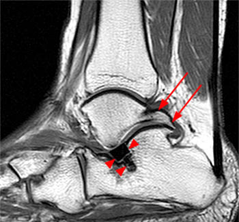
| A sagittal T2-weighted image with fat saturation demonstrates a prominent posterolateral process of the talus (long arrow) with mild edema extending into the talar body (asterisk) and a moderately large volume of surrounding fluid (short arrows). |
 |
| An axial T2-weighted image on the same patient in Figure 7a demonstrates the relationship of the flexor hallucis longus tendon (arrow) to Stieda's process (asterisk). Tenosynovitis of the flexor hallucis longus is also apparent. |
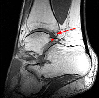 |
| Sag T1-weighted sequence demonstrates a prominent Stieda's process (asterisk) (which showed mild edema on a T2-weighted sequence) and a small dorsal tibial osteophyte (arrow). |
 |
| The sagittal T2-weighted with fat-saturation image demonstrates a prominent os trigonum (asterisk) with surrounding synovitis and mild cyst formation in the corresponding posterior talus (arrow) consistent with chronic posterior impingement. |
 |
| A sagittal T1-weighted image demonstrates prominent osteophyte formation at the posterolateral aspect of the talus and the corresponding calcaneus (arrows). A low signal intensity spacer (arrowheads) is noted in the subtalar joint. Reference |
No comments:
Post a Comment