The constellation of findings that includes synovitis in several compartments of the wrist, an erosion in the distal ulna, tenosynovitis in multiple dorsal extensor tendon compartments, and radial and ulnar flexor bursitis are consistent with rheumatoid arthritis.

Reference

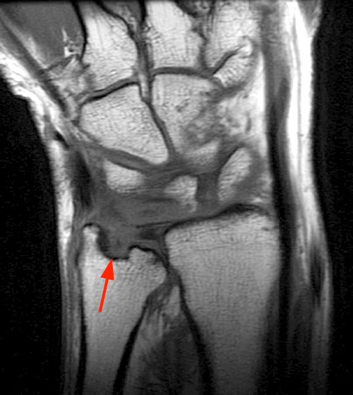 |
| Erosion of the distal ulna |
 |
| The coronal post-contrast image (2b) demonstrates extensive synovitis about the carpal bones, especially in the radiocarpal and midcarpal compartments with additional synovitis in the distal radioulnar joint including the region of the ulnar erosion. |
 |
| The post-contrast axial T1-weighted image with fat suppression at the level of the carpal tunnel (2c) shows evidence of radial and ulnar bursitis surrounding the flexor tendons (arrows) and peritendinous enhancement surrounding the tendons traversing the 1st through 4th dorsal compartments (arrowheads). |
 |
| The axial T2-weighted image with fat suppression (2d) at the level of the distal radioulnar joint demonstrates a focal defect in the subsheath of the extensor carpi ulnaris tendon along its dorsoulnar aspect, consistent with a subsheath injury (arrow). |
Reference
No comments:
Post a Comment