1-Present in the middle of the knee joints.
2-We have two ligaments, anterior and posterior cruciate ligaments.
3-Both originate from posterior surface of the lower femur and then the anterior one inserts in the anterior aspect of the proximal articular surface of the tibia while the posterior one inserts in the posterior part of the proximal articular surface of the tibia.
4-Search first for the posterior cruciate ligament in sagital view as it is large enough to be seen easly, then go one slice medial or lateral to that slice, usually you can see the anterior cruciate ligament if the angle of the technique was taken in a good manner or if the anterior cruciate ligament is not teared, if not, look in the coronal view, you can see the anterior cruciate ligament attached to the lateral femoral condyle in the inter condylar notch if the technique is improper, and if you cannot see it, so it will be teared.
5-Posterior cruciate can be seen with the menisco-femoral ligaments are related to it.
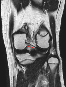 |
| Posterior meniscofemoral ligament on MRI, coronal |
 |
| Posterior meniscofemoral ligament on MRI, sagittal |
 |
| Posterior meniscofemoral ligament (Wrisberg) behind the posterior horn of the lateral meniscus close to its insertion. Sometimes wrongly interpreted as a meniscal tear. |
So in the sagital plane, posterior to the posterior horn of the lateral meniscus, we have two structures which could make confusion with posterior horn tear of the lateral meniscus, these two structures are the tendon of popliteus muscle which appears as vertical linear hypo intense structure posterior to posterior horn, while the second one is posterior menisco-femoral ligament.
 |
| Anterior meniscofemoral ligament(Humphrey ligament) |
No comments:
Post a Comment